Category: New publication

June 20, 2024
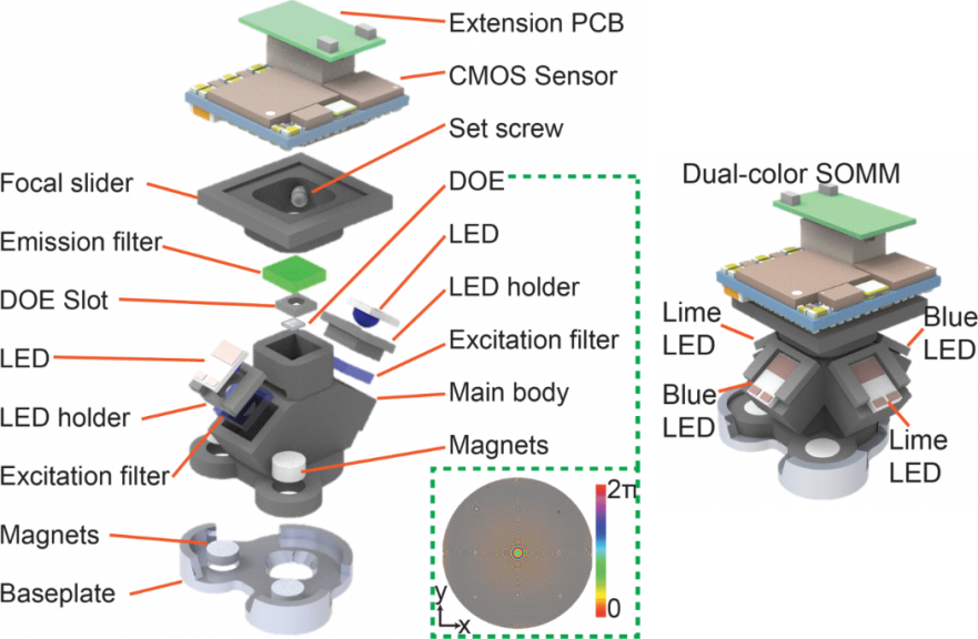
We are happy to announce that a new manuscript entitled “A Systematically Optimized Miniaturized Mesoscope (SOMM) for large-scale calcium imaging…
May 15, 2024
We are excited to announce the publication of our article, “Volatile working memory representations crystalize with practice,” in Nature. This…
April 18, 2024
We are excited to share that our paper entitled "Drugs of abuse hijack a mesolimbic pathway that processes homeostatic need"…
January 16, 2024
We are happy to announce that a new manuscript entitled “Simultaneous, cortex-wide and cellular-resolution neuronal population dynamics reveal an unbounded…
February 23, 2023
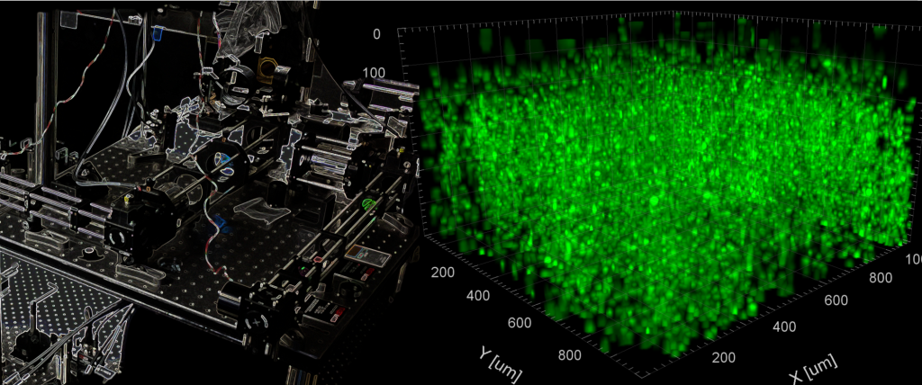
Our article entitled “Mesoscale volumetric light field (MesoLF) imaging of neuroactivity across cortical areas at 18 Hz” has been published in…
March 11, 2021
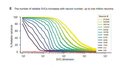
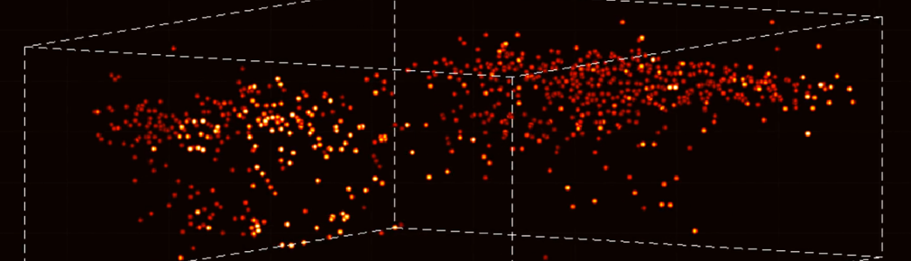
Excited to share our new manuscript showing volumetric Ca imaging of 1 million neurons across the mouse cortex at cellular…
April 11, 2019
Our paper entitled “Volumetric Ca2+ Imaging in the Mouse Brain using Hybrid Multiplexed Sculpted Light (HyMS) Microscopy” has been published…
May 7, 2018
Our paper entitled “High-speed volumetric imaging of neuronal activity in freely moving rodents” has been published in Nature Methods. We…
April 25, 2018
Our review article entitled "A Guide to Emerging Technologies for Large-Scale and Whole-Brain Optical Imaging of Neuronal Activity" has been…March 29, 2018
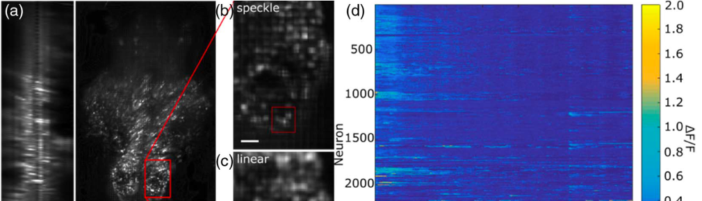
Our article entitled "Brain-wide 3D light-field imaging of neuronal activity with speckle-enhanced resolution" has been published in Optica. We introduce…