January 16, 2024
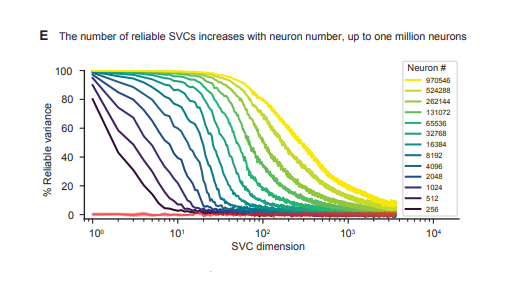
We are happy to announce that a new manuscript entitled “Simultaneous, cortex-wide and cellular-resolution neuronal population dynamics reveal an unbounded…
February 23, 2023
Our article entitled “Mesoscale volumetric light field (MesoLF) imaging of neuroactivity across cortical areas at 18 Hz” has been published in…
October 26, 2022
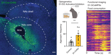
We are happy to share that our paper "Dynamic Processing of Hunger and Thirst by Common Mesolimbic Neural Ensembles" has…
August 30, 2021
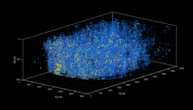
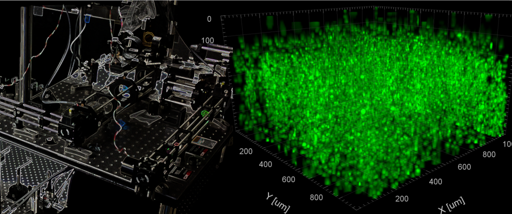
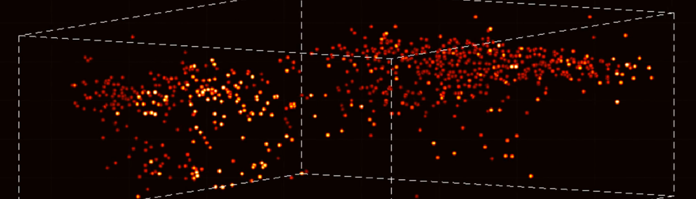
We are excited to share that our paper entitled “High-Speed, Cortex-Wide Volumetric Recording of Neuroactivity at Cellular Resolution using Light…
March 11, 2021
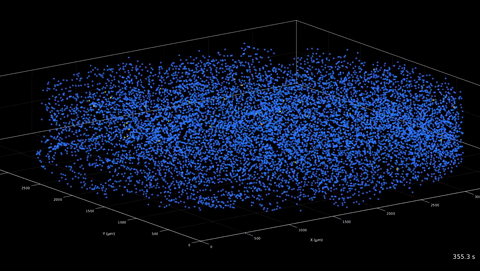
Excited to share our new manuscript showing volumetric Ca imaging of 1 million neurons across the mouse cortex at cellular…
January 16, 2020
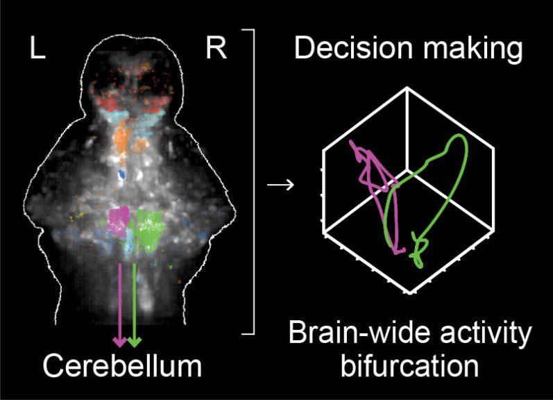
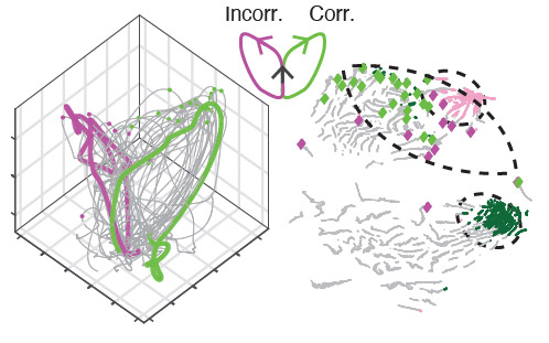
Our paper entitled “Cerebellar neurodynamics predict decision timing and outcome on single-trial level” has been published in Cell. In this…
November 11, 2019
We have uploaded a new manuscript entitled "Cerebellar neurodynamics during motor planning predict decision timing and outcome on single-trial level"…
June 12, 2019
Our recent article in Cell on Hybrid Multiplexed Sculpted Light Microscopy (HyMS) has been featured as a Research Highlight in…April 11, 2019
Our paper entitled “Volumetric Ca2+ Imaging in the Mouse Brain using Hybrid Multiplexed Sculpted Light (HyMS) Microscopy” has been published…
May 7, 2018
Our paper entitled “High-speed volumetric imaging of neuronal activity in freely moving rodents” has been published in Nature Methods. We…