Category: News

March 26, 2026
Optical Systems EngineerPostdoc - Systems Neuroscience Research Assistant
March 26, 2026
The Kavli Fellowship is awarded to graduate students and postdoctoral fellows in the neuroscience community at Rockefeller University, promoting their…
April 18, 2025
Christian received his Ph.D. in Biomedical Engineering from University of Texas at Austin in 2025 where he developed label-free, nonlinear…
December 19, 2024
Jeff received his Ph.D. in Electrical and Computer Engineering from Boston University in 2017 where he worked on Intermodal Parametric…
June 20, 2024
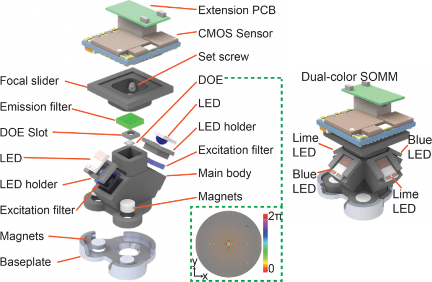
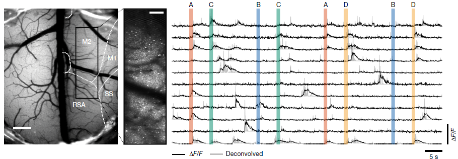
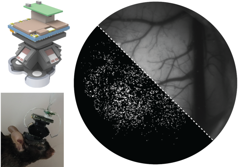
We are happy to announce that a new manuscript entitled “A Systematically Optimized Miniaturized Mesoscope (SOMM) for large-scale calcium imaging…
May 15, 2024
We are excited to announce the publication of our article, “Volatile working memory representations crystalize with practice,” in Nature. This…
April 18, 2024
We are excited to share that our paper entitled "Drugs of abuse hijack a mesolimbic pathway that processes homeostatic need"…
March 26, 2024
Nikita received his Ph.D. in Physics and Mathematics from ITMO University, St. Petersburg, Russia where he worked on structure related…
March 7, 2024
We are happy to announce that a new manuscript entitled “Whole-brain neural substrates of behavioral variability in the larval zebrafish”…
February 23, 2024
We are happy to announce that a new manuscript entitled “A Systematically Optimized Miniaturized Mesoscope (SOMM) for large-scale calcium imaging…