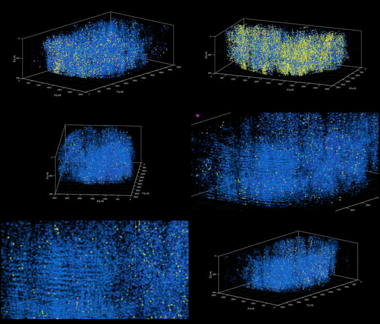
Excited to share our new manuscript showing volumetric Ca imaging of 1 million neurons across the mouse cortex at cellular resolution using Light Beads Microscopy (LBM).

Two-photon microscopy together with genetically encodable calcium indicators has emerged as a standard tool for high-resolution imaging of neuroactivity in scattering brain tissue. However, its various realizations have not overcome the inherent tradeoffs between speed and spatiotemporal sampling in a principled manner which would be necessary to enable, amongst other applications, mesoscale volumetric recording of neuroactivity at cellular resolution and speed compatible with resolving calcium transients. In this paper, we introduce Light Beads Microscopy (LBM), a scalable and spatiotemporally optimal acquisition approach limited only by fluorescence life-time, where a set of axially-separated and temporally-distinct foci record the entire axial imaging range near-simultaneously, enabling volumetric recording at 1.41 × 10⁸ voxels per second.
Read our full publication here.
Congratulations to the entire team!
