
Our paper entitled “Cellular-resolution Three-photon Ultra-Deep Imaging (TrUDI) of neuronal population activity at 2 mm depth in the mammalian brain” has been published in Nature Methods. In this paper we present a systematic optimization approach in three-photon imaging that has enabled cellular resolution calcium imaging of neuronal population activity up to 2 mm depth in the mouse brain.
Three-photon microscopy (3pM) has been shown to extend the depth reach of two-photon microscopy (2pM) in the scattering mammalian brain due to the longer utilized wavelengths that reduce tissue scattering and most importantly by suppressing out-of-focus fluorescence which in 2pM results in loss of contrast, i.e. signal to background ratio (SBR) at a tissue depth of ~ 0.8 mm in the mouse brain (Figure 1a). However, extending the depth reach of 3pM to its fundamental SBR limited regime, which is well beyond 2 mm, remained extremely challenging. The compensation of the exponential loss of in-focus, signal generating photons requires a substantial increase of laser pulse-energy which together with the utilized laser repetition rate needs to remain within limits set by linear and nonlinear safe tissue power exposure (Figure 1b, c). Consequently, previous experimental demonstrations of functional 3p imaging have been limited to depths of about 1.2 mm by either insufficient fluorescence signal at the focus or constraints imposed by laser-induced tissue heating, thus leaving a substantial gap to the theoretically possible depth reach of 3pM.
TrUDI addresses this issue through a bottom-up, systematic optimization of the excitation laser, optical system, and acquisition strategy. It matches the laser repetition rate, pulse energy, pulse duration, excitation point spread function, spatial sampling, field of view, and imaging speed to the requirements of single-cell calcium imaging and to the linear and nonlinear limits of safe laser exposure. All optimization parameters are informed by the required SNR of the baseline fluorescence and discriminability index (d’) for reliable Ca2+ transient detection which determines the required pulse energy (Figure 1d, e). A crucial element of this strategy is single-pulse-per-pixel excitation at a substantially lower laser repetition rate, which maximizes the signal for a given average laser power. This results in a 160 kHz source with ~50 fs pulses at 1300 nm that is used together with a scan engine and optical system that operate at a pixel spacing of ~1.8 µm over a ~230 × 230 µm² field of view at 8.6 Hz. In addition, as we show, at large tissue depths, the highest excitation numerical aperture is not necessarily optimal due to additional losses. TrUDI uses an effective excitation NA of ~0.3 while retaining the full high NA (0.8) of the objective for fluorescence collection (Figure 1f). Through these optimization steps TrUDI realizes an approximately 250-fold increase in three-photon excitation efficiency compared to previous systems based on MHz-rate excitation sources. These optimizations have enabled recordings of neuronal population activity at depths up to ~2 mm in mouse brain tissue while remaining below a verified safe average power level of 150 mW.
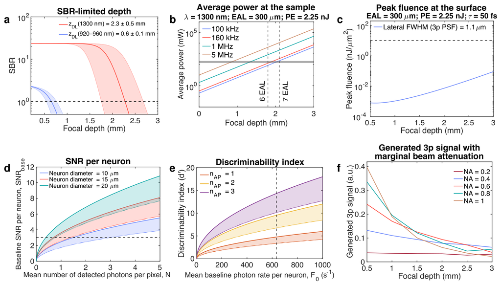
Figure 1: Optimal parameters for background-limited deep-tissue three-photon imaging. (a) Signal-to-background ratio (SBR) as a function of imaging depth in 2pM at 920–960 nm and 3pM at 1300 nm. (b) The required average power for 3pM as a function of imaging depth for different laser repetition rates for a fixed pulse energy (PE) of 2.25 nJ at the focus. (c) The peak fluence at the surface as a function of focal depth. (d) Baseline SNR per neuron as functions of the mean number of detected photons per pixel at the PMT. (e) Discriminability index (d’), a measure of neuroactivity SNR, as a function of mean baseline photon rate per neuron for different numbers of action potentials. (f) Generated 3p fluorescence signal as a function of focal depth for different excitation NAs.
We demonstrate the capabilities of our imaging system by performing imaging throughout the layers of the motor cortex and into the striatum, as well as through the posterior parietal cortex (PPC) into the various regions of the hippocampus, including the CA1 layer, the CA3 layer, and the dentate gyrus (DG) in awake mice. Figures 2, 3 demonstrate examples of cellular resolution calcium imaging at depths up to 2 mm in these brain regions in the mouse brain which previously remained inaccessible.
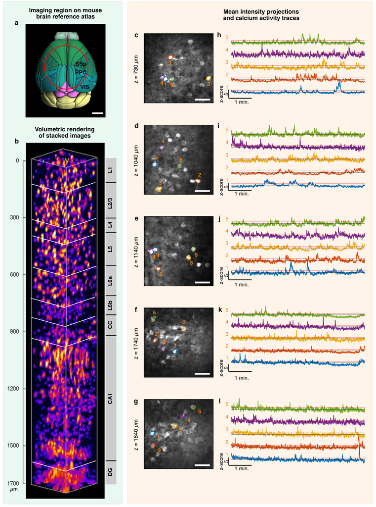
Figure 2: TrUDI enables deep tissue calcium (Ca2+) imaging over a ~1800 µm depth range, allowing imaging of the majority of hippocampus (HPC) layers through intact posterior parietal cortex (PPC) in mice expressing GCaMP8m and GCaMP8s. (a) Approximate location of an 8-mm cranial window over the PPC and the subcortical HPC on Allen mouse brain atlas. (b) High-resolution reconstructed volume stack covering a ~1700 µm depth range, showing neurons at different layers (L1–L6) of the PPC, as well as hippocampal CA1 and DG region of mouse expressing GCaMP8m (VGAT-Cre × GCaMP8m). (c-g) Mean intensity projection of neuroactivity in individual planes covering a depth range of 730–1840 µm in mouse brain expressing GCaMP8s (Camk2a-tTA × tetO-GCaMP8s). The maximal average power used for the deepest layer was 66 mW, which was substantially lower than the established upper bound for safe power limits. (h-l) Ca2+ activity traces of selected neurons numbered in (c-g) over ~6 min. Traces were extracted from recordings using CaImAn with z-scores up to ~ 10 for the strongest responding neurons.
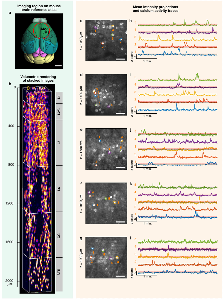
Figure 3: TrUDI enables deep-tissue calcium (Ca2+) imaging over a ~2000 µm depth range within motor cortical areas and the striatum of mice expressing GCaMP6s. (a) Approximate location of a 5-mm cranial window over the mouse brain, targeting deep imaging in the secondary motor cortical area (M2) and striatum, shown over the Allen mouse brain atlas. (b) High-resolution reconstructed volume stack covering a ~2060 µm depth range, showing neurons at different layers (L1–L6) of M2 as well as in the striatum for mouse expressing GCaMP6s (Camk2a-tTA × tetO-GCaMP6s). The acquisition power was exponentially increased from 0.5 to 150 mW with depth. (c-g) Mean intensity projection of neuroactivity in individual planes covering a depth range of 1050–1930 µm in mouse brain expressing GCaMP6s. The maximal average power used for the deepest layer was 120 mW. (h-l) Ca2+ activity traces of selected neurons numbered in (c-g) over ~ 4 min. Traces were extracted from recordings using CaImAn and show z-scores up to ~12 for the strongest responding neurons.
Finally, we demonstrate that TrUDI can further reduce average power by avoiding excitation of image pixels that do not contribute useful neuronal signal, which can be achieved by suppressing laser pulses outside the selected regions using a fast electro-optic modulator. In an example at 1.14 mm depth, this targeted-excitation strategy reduced average laser power to approximately 18–28% of the full-field illumination level while maintaining comparable calcium-activity z-scores.
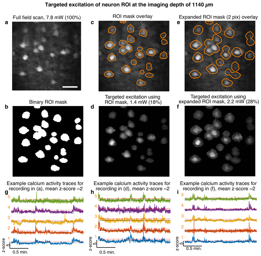
Figure 4: Targeted excitation minimizes laser exposure in TrUDI. (a) A representative full-field scanned image at 1140 µm depth using an average power of 7.8 mW. (b) Segmented neuronal regions-of-interest (ROIs) binary mask generated for the image shown in (a). (c) Overlay of binary ROI mask extracted in (b) with image (a) resulting in a pixel coverage of ~18% of the area of the full field image. (d) Image acquired with power modulation over the same field-of-view (FOV) as in (a) using the ROI mask overlay shown in (c) resulting in a total average power of 1.4 mW, i.e. ~ 18% of that used for full field imaging. (e) Overlay of expanded ROI by 2 pixels along the perimeter of the ROIs extracted in (b) resulting in a pixel coverage of ~28% of the area of the full field image. (f) Image acquired with power modulation over the same FOV as in (a) using the expanded ROI mask overlay shown in (e) resulting in a total average power of 2.2 mW, i.e. ~ 28% of that used for full field imaging. (g-i) Ca2+ activity traces of representative neurons for the recordings in (a), (d), and (f) are shown in (g), (h), and (i), respectively. Traces were extracted from recordings using CaImAn after motion correction. The mean z-score in all three recordings is ~ 2.
The above results demonstrated the capabilities of TrUDI which are expected to open up new possibilities for studying cellular-resolution dynamics of neuronal populations across a tissue column at previously unavailable depths within the scattering mouse brain.
Relevant Publication:
K. Santra, K. Barber, F. Logiacco, M. A. Frechou, A. Bellafard, J. Liou and A. Vaziri, Cellular-resolution Three-photon Ultra-Deep Imaging (TrUDI) of neuronal population activity at 2 mm depth in the mammalian brain, Nature Methods (2026), doi:10.1038/s41592-026-03251-2
https://www.nature.com/articles/s41592-026-03251-2?utm_source=chatgpt.com